
Supplier:
United States BiologicalCat no: 031368
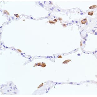
Glial Fibrillary Acidic Protein (GFAP)
Prices direct from United States Biological
Quick response times
Exclusive Biosave savings/discounts
SPECIFICATIONS
Catalog Number
031368
Size
100ul
Applications
IF, IHC, WB
Hosts
Mouse
Reactivities
Hum, Mouse, Rat, Bov, Prc
Form
Supplied as a liquid.
P Type
Mab
Purity
Ascites
Isotype
IgG1,k
References
1. Bignami A, Eng LF, Dahl D, Uyeda CT. Localization of the glial fibrillary acidic protein in astrocytes by immunofluorescence. Brain Res. 43:429-35 1972.\n\n2. Yen SH, Fields KL. Antibodies to neurofilament, glial filament, and fibroblast intermediate filament proteins bind to different cell types of the nervous system. J Cell Biol. 88:115-26 1981.\n\n3. Shaw G, Osborn M, Weber K. An immunofluorescence microscopical study of the neurofilament triplet proteins, vimentin and glial fibrillary acidic protein within the adult rat brain. Eur J Cell Biol. 26:68-82 1981.\n\n4. Fitch MT, Silver J. CNS injury, glial scars, and inflammation: Inhibitory extracellular matrices and regeneration failure. Exp Neurol. 209:294-301 2008.\n\n5. Brenner M, Johnson AB, Boespflug-Tanguy O, Rodriguez D, Goldman JE, Messing A. Mutations in GFAP, encoding glial fibrillary acidic protein, are associated with Alexander disease. Nat Genet 27:117-20 2001.
Additional Info
Recognizes GFAP from human, bovine, porcine, equine, mouse, rat and all other mammalian and avian species tested to date. It is strong and clean on western blots and works well on frozen sections, cells in tissue culture and on formalin fixed histological sections.
SUPPLIER INFO


Applications
ELISA
Reactivities
Hum
Applications
IF
Hosts
Mouse
Applications
ELISA, WB
Hosts
Mouse
Reactivities
Hum
Applications
ELISA, FC, WB
Hosts
Mouse
Reactivities
Hum
Applications
ELISA, FC, IHC, WB
Hosts
Mouse

Latest promotions
Spend less time on DNA cleanup so you can do more science. The MSB Spin PCRapace is the fastest way to purify your DNA from PCR, restriction digestion, and...
New brilliant antibodies, and new lower prices!For flow cytometry reagents in general, \"bright is better.\" The violet-excitable BD Horizon™ BV421 and...
As an incentive to qualify our BSA, we are offering a 20% discount when you purchase your first 100g, 500g or 1000g of any grade of Bovine Serum Albumin....
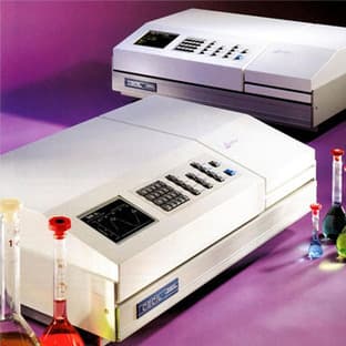
It is not every day that you are given something for nothing. We are giving away additional spectrophotometer software.Cecil Instruments have enhanced the...
Did your supplier increase the price of Fetal Bovine Serum? Did they substitute the US Origin with USDA? Well say no more! Innovative Research is still...
We're so sure that you'll prefer Cayman Assay kits over your present brand that we're willing to give you a free assay kit to prove it!

For the past decade scientists have extensively used ATS secondary toxin conjugates to make their own targeted toxins for in vitro use.The ability to combine...
10% Discount on 2 Rabbit Polyclonal Antibody Service. With over 20 years experience, SDIX has developed into the premier US custom antibody producer,...
Bulk Cytokines with Custom Vialing.20 - 50% off cytokines, growth factors, chemokines and more...For a limited time Cell Sciences is offering substantial...
Are you planning to have a customised antibody made for your research?Since 2000, Everest has been producing a catalog containing thousands of affinity...
Jenway’s 73 series spectrophotometer range provides four models with a narrow spectral bandwidth of 5nm and an absorbance range of –0.3 to 2.5A,...
Top suppliers

United States Biological
230747 products
Carl Zeiss Microscopy
27 products
Promega Corporation
11 products
Panasonic Healthcare Company
5 products

Life Technologies
1 products

Nikon Instruments Europe
11 products

Olympus Europa Holding GmbH
3 products
Leica Microsystems, Inc.
10 products

GE Healthcare Life Sciences
2 products

Tecan Trading AG
19 products

Beckman Coulter, Inc.
1 products

AB SCIEX
3 products

BD (Becton, Dickinson and Company)
1 products
RANDOX TOXICOLOGY
5 products

Randox Food Diagnostics
6 products