
Supplier:
United States BiologicalCat no: V2122-03A
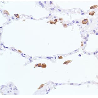
Vimentin (FLJ36605, VIM)
Prices direct from United States Biological
Quick response times
Exclusive Biosave savings/discounts
SPECIFICATIONS
Catalog Number
V2122-03A
Size
50ug
Applications
ELISA, IF, IHC, WB
Hosts
Mouse
Reactivities
Hum, Bov, Can, Ch/Bird, NHP
Form
Supplied as a lyophilized powder in PBS, pH 7.4, 0.5% BSA, 0.09% sodium azide. Reconstitute to a final volume of 1ml.
P Type
Mab
Purity
Purified by Protein A affinity chromatography.
Isotype
IgG2a
References
Heid HW, Moll I, Franke WW: Patterns of expression of trichocytic and epithelial cytokeratins in mammalian tissues I: Human and bovine hair\nfollicles. Differentiation 37, 137-157 (1988)\nJahn L, Fouquet B, Rohe K, Franke WW: Cytokeratins in certain endothelial and smooth muscle cells of two taxonomically distant vertebrate\nspecies, Xenopus laevis and man. Differentiation 36, 234-254 (1987)\nKasper M, Stosiek P, van Muijen GNP, Moll R: Cell type heterogeneity of intermediate filament expression in epithelia of the human pituitary\ngland. Histochemistry 93, 93-103 (1989)\nKasper M, Karsten U, Stosiek P, Moll R: Distribution of intermediate-filament proteins in the human enamel organ: Unusually complex pattern of\ncoexpression of cytokeratin polypeptides and vimentin. Differentiation 40, 207-214 (1989)\nMoll I and Moll R: Comparative cytokeratin analysis of sweat gland ducts and eccrine poromas. Arch Dermatol Res 283, 300-309 (1991).\nGomi H, Yokoyama T, Fujimoto K, Ikeda T, Katoh A, Itoh T Itohara S: Mice Devoid of the Glial Fibrillary Acidic Protein Develop Normally and Are\nSusceptible to Scrapie Prions. Neuron, 14, 29-41 (1995)\nDemirkesen C, Hoede N, Moll R: Epithelial markers and differentiation in adnexal neoplasms of the skin: an immunohistochemical study\nincluding individual cytokeratins. J Cutan Pathol 22: 518-535 (1995).\nHermann H, Eckelt A, Brettel M, Grund C, Franke WW: Temperature-sensitive intermediate filament assembly. Alternative structures of\nXenopus laevis vimentin in vitro and in vivo. J Mol Biol 234: 99-113 (1993).\nRogers KR, Eckelt A, Nimmrich V, Janssen K-P, Schliwa M, Hermann H, Franke WW: Truncation mutagenesis of the non-a-helical\ncarboxyterminal tail domain of vimentin reveals contributions to cellular localization but not to filament assembly. Eur J Cell Biol 66: 136-150\n(1995).\nBohn W, Wiegers W, Beuttenm
Additional Info
Recognizes the intermediate filament protein vimentin at 57kD, which is present in all cells of mesenchymal origin. Species Crossreactivity: Human, monkey, bovine, canine, chicken, amphibia.
SUPPLIER INFO


Applications
ELISA, FC, IHC, WB
Hosts
Mouse
Applications
IHC, WB
Hosts
Rabbit
Reactivities
Hum
Applications
ELISA, WB
Hosts
Rabbit
Reactivities
Hum
Applications
ELISA
Hosts
Mouse
Reactivities
Hum, Mouse
Applications
ELISA
Hosts
Mouse
Reactivities
Hum, Mouse
Applications
ELISA
Hosts
Mouse

Latest promotions

Spend less time on DNA cleanup so you can do more science. The MSB Spin PCRapace is the fastest way to purify your DNA from PCR, restriction digestion, and...
New brilliant antibodies, and new lower prices!For flow cytometry reagents in general, \"bright is better.\" The violet-excitable BD Horizon™ BV421 and...
As an incentive to qualify our BSA, we are offering a 20% discount when you purchase your first 100g, 500g or 1000g of any grade of Bovine Serum Albumin....
It is not every day that you are given something for nothing. We are giving away additional spectrophotometer software.Cecil Instruments have enhanced the...

Did your supplier increase the price of Fetal Bovine Serum? Did they substitute the US Origin with USDA? Well say no more! Innovative Research is still...

For the past decade scientists have extensively used ATS secondary toxin conjugates to make their own targeted toxins for in vitro use.The ability to combine...
We're so sure that you'll prefer Cayman Assay kits over your present brand that we're willing to give you a free assay kit to prove it!

10% Discount on 2 Rabbit Polyclonal Antibody Service. With over 20 years experience, SDIX has developed into the premier US custom antibody producer,...
Bulk Cytokines with Custom Vialing.20 - 50% off cytokines, growth factors, chemokines and more...For a limited time Cell Sciences is offering substantial...
Jenway’s 73 series spectrophotometer range provides four models with a narrow spectral bandwidth of 5nm and an absorbance range of –0.3 to 2.5A,...
Are you planning to have a customised antibody made for your research?Since 2000, Everest has been producing a catalog containing thousands of affinity...
Top suppliers

United States Biological
230747 products
Carl Zeiss Microscopy
27 products
Promega Corporation
11 products
Panasonic Healthcare Company
5 products

Life Technologies
1 products

Nikon Instruments Europe
11 products

Olympus Europa Holding GmbH
3 products
Leica Microsystems, Inc.
10 products

GE Healthcare Life Sciences
2 products

Tecan Trading AG
19 products

Beckman Coulter, Inc.
1 products

AB SCIEX
3 products

BD (Becton, Dickinson and Company)
1 products
RANDOX TOXICOLOGY
5 products

Randox Food Diagnostics
6 products